‘Noon’ means EYE in Korean.
SNU Eye Clinic will be with you
On your journey to regain your natural vision

scroll down

SNU EYE CLINIC

Restore the lost brightness of eyesight
Regain the original bright vision

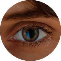
Our eyeballs resemble the phases of the moon.
The moon changes from a full moon to a crescent moon over time due to its orbit.
Likewise, people gradually lose their original vision for different reasons.

SNU Eye Clinic believes that vision correction is not just a simple surgery,
but a journey to regain everyone’s original shine.

Just as the crescent moon regains its light and returns to a full moon,
SNU Eye Clinic will be with you on your journey to regain your original bright vision.

SNU MEDICAL STAFF
Chung Eui Sang Chief Director

Introduction of SMILE Lasik in Korea,
first SMILE Eye Surgery and Toric ICL surgery 
in Korea, initial clinical research in progress

  • Seoul National University Graduate School, Doctor of Medicine
  • Full-time doctor at Seoul National University Hospital
  • Full-time doctor at Samsung Seoul Hospital
  • Professor, Samsung Seoul Hospital, Sungkyunkwan University School of Medicine
  • Director of Daejeon Eye Hospital
  • Havard Schepens Eys Research Institute Research Professor
  • SMILE Certified Doctor
  • TICL Reference Doctor
  • ICL &TICL EXPERT SURGEON
SNU MEDICAL STAFF
Kim Joo Hyun Director

Patient-tailored vision correction that takes into account the axis of astigmatism

  • Bachelor of Medicine, Hallym University College of Medicine
  • Completed Master of Medicine degree from Hallym University Graduate School
  • Hallym University Gangnam Sacred Heart Hospital Intern
  • Best resident at Hallym University Gangnam Sacred Heart Hospital
  • Full-time doctor, Department of Ophthalmology, Seoul National University Bundang Hospital
  • Director of Seoul Bright World Ophthalmology Clinic
  • Smile Certified Doctor
  • ICL &TICL EXPERT SURGEON
SNU MEDICAL STAFF
Lee Min Gyu Director

Graduated as the best doctor at University Hospital
1:1 Customized Vision Correction

  • Bachelor of Medicine, Sungkyunkwan University School of Medicine
  • Samsung Seoul Hospital trainee
  • Ophthalmology resident at Samsung Seoul Hospital (top resident)
  • Director of Seoul Samsung Eye Clinic
  • Director of Bright St. Mary’s Eye Clinic Gangnam
  • SMILE Certified Doctor
  • SMART LASIK certified doctor
SNU MEDICAL STAFF
Kim Seong Hee Director

Operation of a postoperative observation system

  • Bachelor of Medicine, Seoul National University College of Medicine
  • Seoul Asan Hospital trainee
  • Resident at Samsung Seoul Hospital
  • Samsung Seoul Hospital specialist
  • Department of Ophthalmology, Armed Forces Yangju Hospital
  • Chief of Ophthalmology, Jeonju Hospital
  • Director of Mokpo Seoul Ophthalmology Clinic
  • Adjunct Professor, Sungkyunkwan University School of Medicine
SNU EYE CLINIC

SNU Ophthalmology Optimized for
Global Patients

Available staff to
accommodate foreigners.
A total of 1,200 global patients visited SNU Eye Clinic between 2022 and 2023.
More than 1,000 global patients who underwent vision correction surgery between 2022 and 2023.
SMILE LASIK

SMILE Eye Surgery
there is SNU Eye Clinic
on the beginning

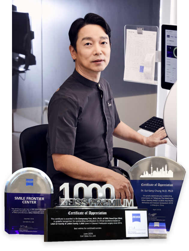
One of 11 International Smile Research Doctors, CEO Chung Eui-sang
In May 2011, Smile LASIK was introduced domestically and initial clinical research was conducted.
Leading understanding of Visumax equipment and surgical methods using femtosecond lasers
ZEISS Officially Certified Ophthalmology Clinic
Designated as a frontier center leading SMILE
All medical staff are officially certified doctors by ZEISS SMILE

Why is SMILE Lasik in SNU Eye Clinic Special?

HISTORY

2006

First Visumax Development

2008

European CE Approval

2009

Approved by the domestic Ministry of Food and
Drug Safety

2010

Visit to Carl Zeiss headquarters in Germany, tour of Visumax equipment production process and meeting with CEO, later visits to Germany and India, observation of SMILE surgery and transfer of surgical techniques

2011 May

Director Chung Eui Sang performed SMILE surgery and gave clinical presentations while working at Samsung Seoul Hospital.

2011

SMILE Korean Clinical Results Presented at International Conference: 2011 APCRS (Asia Pacific Cataract and Refractive Surgery Society)

2012

SMILE clinical results presented at International Academic Conferences: APAO (Asian Pacific Association of Ophthalmologists), 2012 ESCRS (European Society of Cataract and Refractive Surgery)

2018

Approved by US FDA

2020

SNU SMILE Clinic Extension

2021

Visumax 21 Model Extra Introduction

2022

Reached 1 Million in Korea Reached 7 Million Worldwide

2023 Aug 14th

Introduction of SMILE Pro in SNU Eye Clinic and First Surgery

Characteristic of SMILE Eye Surgery

Surgery that makes a minimal incision of only 2mm in the cornea
Instead of cutting or opening the surface of the cornea, the laser is used to separate only the parenchymal area by the amount of correction.
Femtosecond laser SMILE Eye Surgery – Pain relief through quick surgery, quick return to daily activities.
600 times lower energy sophistication.Enhanced stability
High myopia and high astigmatism can be corrected, minimizing the probability of myopic regression

Eyes suitable for SMILE Eye Surgery

CASE 01High myopia
CASE 02High astigmatism
CASE 03If the pupil is large
CASE 04If there is a
corneal disease
CASE 05Corneal asymmetry
CASE 06If dryness is severe

SNU Eye Clinic adjusts

the size of the optical unit, cutting amount, axis of astigmatism,
and energy irradiation according to the characteristics of each patient.

SMILE PRO

Smile Pro, the choice of professionals for perfect results

Innovative technology

Extremely accurate.

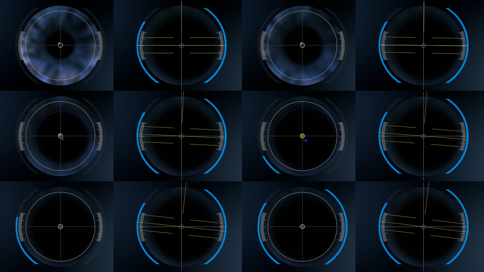
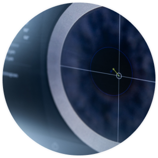
  • CentraLign

    It accurately detects eye movements and
    automatically takes the center.

  • OcuLign

    By correcting the rotation of the astigmatism axis,
    more complete surgery is possible.

Faster.

More than 3x faster laser exposure

Robotic.

Smart robotic assistance, easy patient access
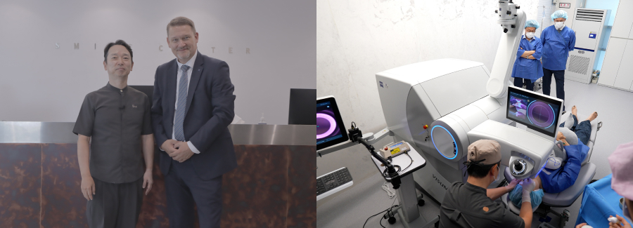
Smile Pro preemptively introduced clinic
to achieve 100 eye surgeries
for the first time in Korea

The general manager of refractive surgery
at ZEISS headquarters in Germany
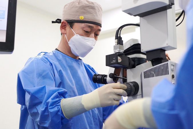
visits the first Smile Pro surgery site in Korea.

Features of Smile Pro

  • Live monitoring of surgical process
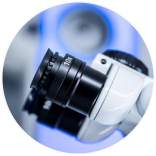
  • ZEISS ultra-precision ophthalmic surgical microscope
  • 2 million laser irradiations per second Laser irradiation time reduced to only 7 seconds
  • Equipped with an automatic pressure compensation system that automatically corrects even the slightest movement of the patient
SNU EYE CLINIC

SNU’s unique bespoke smile for your eyes.

Eyes appropriate for Smile Pro

CASE 01High myopia
CASE 02High astigmatism
CASE 03Corneal asymmetry
CASE 04Those with a strong
fear of surgery
CASE 05Those who want faster recovery and clear vision results
LENS IMPLANTATION SURGERY

Accurate eye measurement, unmatched know-how
for over
20 years of lens implantation

Customized lens insertion optimized for each patient’s eye space SNU FITTING

Lens implantation surgery at SNU Eye Clinic is safer because it uses 3D equipment to structurally identify the eye, determine the type of lens
and insertion location appropriate for the individual, and then perform the surgery.

Chung Eui Sang
  • 01Lens implantation surgery has been performed since the introduction of lens implantation in Korea in 2001.

  • 02Toric ICL lens turnover rate is less than 1%
    (statistics for patients with astigmatism correction lens implantation at SNU Ophthalmology)

  • 032005 Toric ICL (astigmatism correction posterior lens implantation) domestically introduced and clinical research conducted

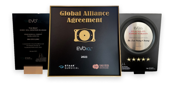
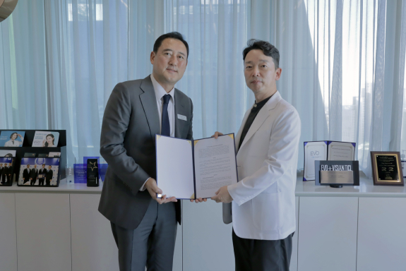
  • 04Agreement ceremony between EVO ICL manufacturer STARR
    and Excellent Lens Implantation Hospital

Lens implantation surgery method

Unlike vision correction surgery using lasers, the cornea is preserved without being cut.
This is a surgical method to correct vision by inserting lenses of a certain capacity.

Appropriate eye for
lens implantation surgery

CASE 01When LASIK and LASEK are not possible
CASE 02When the cornea is too thin compared to the refractive error
CASE 03When the pupil size is large at night
CASE 04If you have severe dry eye syndrome
CASE 05If you have a corneal disease such as keratoconus
PRESBYOPIA, CATARACT

Who would you entrust to your only eye for Presbyopia Cataract Surgery?

Ophthalmologist who has undergone
presbyopia cataract surgery before will perform the surgery.

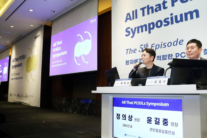
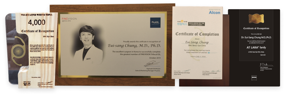
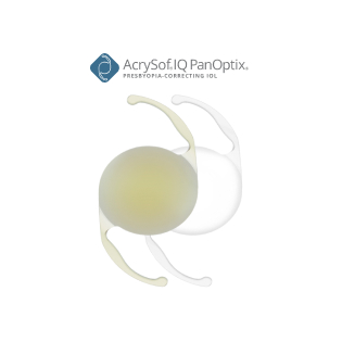
Reference doctor Chung Eui Sang of multifocal intraocular lens company Alcon's panoptics and BBTI lenses

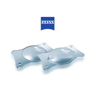
Alcon, ZEISS, Fine Vision, etc.
Various multifocal intraocular lenses.
Recognized as a reference doctor.

Presbyopia symptoms that become
invisible as you age VS Cataracts in presbyopia
usually come together.

  • Presbyopia

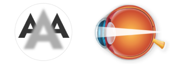
    As one ages, there is a phenomenon where nearby objects are not seen well, due to a decrease in the elasticity of the eye's lens and an weakening of the ability to adjust its thickness.

  • Cataract

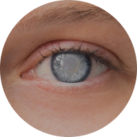
    It is a disease where the lens becomes cloudy, similar to frosted glass, leading to a decline in vision. We recommend seeking treatment before the vision impairment worsens.

Choosing the most appropriate surgical method based on precise examinations is crucial,
as symptoms and conditions can vary among patients.

Multifocal Intraocular Lens

Multifocal intraocular lens implantation surgery that can correct presbyopia and cataracts at once.

SNU Eye Clinic recommends the most suitable lens for the patient based on information such as
the patient's vision, disease status, lifestyle, and primary field of view.

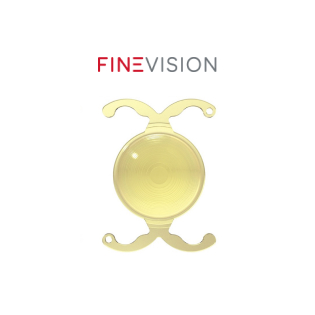
Finevision
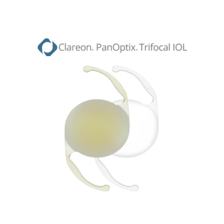
Panoptics
Eyhance
Lisatree Family
Lara Lens
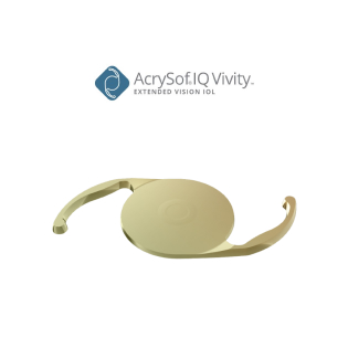
Vivit
Clareon Lens
HIGH TECHNOLOGY EQUIPMENT

We provide customized treatment to
each patient through
52 detailed examination programs.

Experience safer examination and surgical results by introducing high-tech equipment.

Vision Correction Surgical Equipment

zoom_inmoreVisuMax

VisuMax

VisuMax

VisuMax

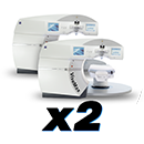
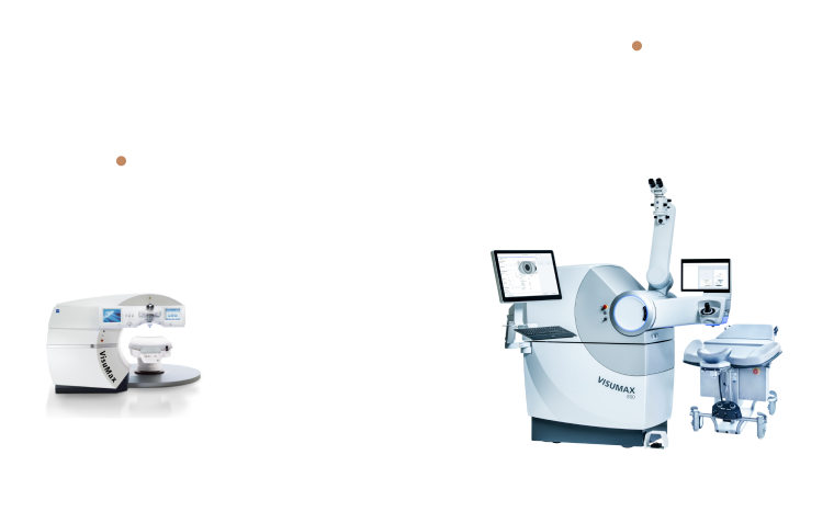
‘Smile LASIK’ surgical equipment for vision correction using a femtosecond laser!

SNU Eye Clinic has a total of 2 Smile Surgery Equipment, including the 2021 Visumax Model.
zoom_inmoreMel90

Mel90

Mel90

Mel90

This is ZEISS' excimer laser equipment, which is a laser device that can perform minimal cutting of the cornea. Mel90 allows for safer LASIK surgery by maximizing corneal thickness.
zoom_inmoreLumera700

Lumera700

Lumera700

Lumera700

A surgical microscope from Germany's ZEISS that allows for fast and accurate surgery!

It is a state-of-the-art surgical microscope that boasts excellent surgical field clarity and high stability.
zoom_inmoreCallisto eye

Callisto eye

Callisto eye

Callisto eye

Equipment that serves as a navigation device that allows for more precise surgery!

The microscope's visual navigation function, including the location and extent of incision and the axis of astigmatism, makes surgery safer and more accurate.
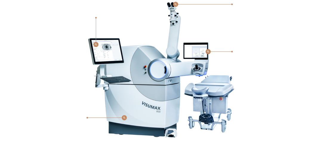
zoom_inmoreVisuMax 800

VisuMax 800

VisuMax 800

VisuMax 800

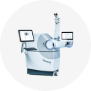
It was introduced domestically in the fall of 2023 as an upgraded surgical equipment optimized for patients with high myopic astigmatism, with safer and faster laser irradiation time and minimal suction loss probability from the existing smile surgery equipment, VisuMax.

This is the Smile Pro surgical equipment that was preemptively introduced to about 10 ophthalmology clinics across the country.

Cataract Surgical Equipment

zoom_inmoreCatalys Precision Laser

Catalys Precision Laser

Catalys Precision Laser

Catalys Precision Laser

State-of-the-art cataract surgery equipment with proven safety and high surgical success rate!

This laser is dedicated to cataract surgery, created through joint research by world-class cataract experts. It is a state-of-the-art cataract surgery laser equipment that complements the shortcomings of general manual surgery.
zoom_inmoreCenturion

Centurion

Centurion

Centurion

Safer and faster artificial intelligence cataract surgery equipment

It automatically maintains intraocular pressure during surgery and maximizes optic nerve protection and stability, enabling rapid vision recovery and minimizing pain during surgery.

Retinal surgical equipment

zoom_inmoreConstellation

Constellation

Constellation

Constellation

The best retina surgery equipment to minimize surgical complications and vision loss!

It is the best retina surgery equipment with a fast cutting speed of 7500rpm, stable intraocular pressure maintenance, and simultaneous cataract/retina surgery.

Optometry equipment

zoom_inmoreCASIA 2

CASIA 2

CASIA 2

전안부 촬영장비

CASIA 2

전안부 촬영을 기존보다 더 깊이(13mm), 더 빠른 속도(0.3초)로 스캔합니다. 1마이크로미터 단위로 360도 촬영하여 다양한 각도에서 고해상도로 각막 지형 및 IOL시뮬레이션이 가능한 장비입니다.
zoom_inmoreK5M Keratograph 5M Tear Film Analysis Test

K5M Keratograph 5M Tear Film Analysis Test

K5M Keratograph 5M Tear Film Analysis Test

K5M Keratograph 5M Tear Film Analysis Test

K5M Keratograph 5M Tear Film Analysis Test

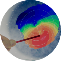
Equipment that accurately analyzes the current condition by determining the movement of the fat layer and moisture layer, tear layer thickness, meibomian glands, tear depth, corneal hyperemia, tear film destruction time, etc.
zoom_inmorePENTACAM

PENTACAM

PENTACAM

PENTACAM

This is a 3D anterior segment CT imaging equipment that photographs and analyzes the anterior segment of the eye, including the front and back of the cornea, anterior chamber, and lens.
zoom_inmoreSpecular Microscope

Specular Microscope

Specular Microscope

Specular Microscope

This is a corneal endothelial cell tester that tests for damage to the corneal endothelial tissue before and after surgery.
zoom_inmoreARK

ARK

ARK

ARK

An automatic refractive power test device measures the refractive value, power, and corneal curvature of the eye.
zoom_inmoreIOP

IOP

IOP

IOP

This is a device that tests intraocular pressure and corneal thickness.

If intraocular pressure is not maintained well, diseases such as optic nerve damage, glaucoma, and keratoconus may occur.
zoom_inmoreOCT Optovue

OCT Optovue

OCT Optovue

OCT Optovue

This is a device that images the retina and optic nerve, and can accurately and quickly examine the retina from the vitreous to the choroid.
zoom_inmoreHumphrey® Matrix

Humphrey® Matrix

Humphrey® Matrix

Humphrey® Matrix

This is a test machine that can detect the early damage of glaucoma, shortening the time required for a typical visual field test, allowing accurate test results to be confirmed in a shorter period of time.
zoom_inmoreF/P

F/P

F/P

F/P

This is a fundus imaging examination device that can check for signs of abnormalities such as the retina and optic nerve, and quickly and simply photographs the condition of the retina using a special fundus camera.
zoom_inmoreGalilei G4

Galilei G4

Galilei G4

Galilei G4

Corneal topography covers everything from basic eye movement compensation to wavefront analysis!

State-of-the-art equipment essential for successful cataract surgery and vision correction surgery. It obtains 60 images per second, obtains information from up to 100,000 measurement points, and provides corneal topography and corneal tomography information at a level higher than existing equipment, enabling sophisticated cataract surgery and vision correction surgery.
zoom_inmoreMovu Argos

Movu Argos

Movu Argos

Movu Argos

The world's first advanced equipment to accurately and successfully measure the eye using SS-OCT.

A comfortable device that is non-invasive and non-contact. This is a test equipment that precisely measures the axial length of the eye to determine the power of the lens to be used during cataract surgery. The test is performed in a non-contact manner within a short period of time. In particular, it boasts excellent performance as it measures with high accuracy even in severe cataracts.
zoom_inmoreKowa nonmyd WX 3D

Kowa nonmyd WX 3D

Kowa nonmyd WX 3D

Kowa nonmyd WX 3D

Equipment that can capture high-quality optic nerve and retina images at the same time even if the pupil size is small

A closer look to reality through 3D image reconstruction. It is an essential examination equipment for retina and glaucoma treatment because it can obtain high-quality images without mydriasis and reconstructs and displays 3D images.
zoom_inmoreKONAN NSP-7700

KONAN NSP-7700

KONAN NSP-7700

KONAN NSP-7700

Complete cell analysis in seconds High reproducibility of similar results even when measured multiple times Essential equipment for safe surgery

It is a precision measuring device used to analyze the condition of corneal endothelial cells, which is difficult to detect with the naked eye.
zoom_inmoreKAXIN ODU8

KAXIN ODU8

KAXIN ODU8

KAXIN ODU8

Fast measurement speed of 30 frames per second with measurement error within 0.02mm

Equipment for measuring axial length, anterior chamber/lens thickness, etc. This is an equipment used to diagnose intraocular diseases when the intraocular condition cannot be accurately determined due to media opacity, or to measure anterior chamber/lens thickness, axial length, etc.
zoom_inmoreDRI OCT-Atlantis

DRI OCT-Atlantis

DRI OCT-Atlantis

DRI OCT-Atlantis

Equipment that shows outstanding precision by accurately visualizing the vitreous body and choroid at an incredible speed of 100,000 scans per second

Equipment that shows outstanding precision by accurately visualizing the vitreous body and choroid at an incredible speed of 100,000 scans per second
zoom_inmoreCirrus 6000

Cirrus 6000

Cirrus 6000

Cirrus 6000

Ocular optical tomography

CRRUS ® 6000 is the next generation of OCT from ZEISS, offering high-speed image capture with HD imaging detail and a wider field of view. With 100,000 scans per second, it is possible to capture images with a wide field of view of up to 12mm in one shot, and the high-resolution octet allows you to see detailed information about the microvessels of the retina.
zoom_inmoreOptos Da

Optos Da

Optos Da

Optos Da

Expanding the shooting range from the existing 50 degrees to 200 degrees

State-of-the-art equipment that can obtain images of the periphery of the retina in less than 1 second without mydriasis. Supports color, autofluorescence, and fluorescein angiography modes. It is a cutting-edge device that reduces discomfort but increases convenience by enabling imaging of the periphery of the retina at once without mydriasis. Retina imaging equipment.
zoom_inmoreHumphrey field analyzer

Humphrey field analyzer

Humphrey field analyzer

Humphrey field analyzer

Precision equipment that can quickly, accurately and easily inspect the field of view

Equipped with GPA (Guided Progression Analysis), it allows early detection of visual field abnormalities. It is an essential equipment that is the gold standard for visual field in the treatment of glaucoma. It is a device used to determine visual field abnormalities and is a state-of-the-art test equipment that boasts high accuracy.
zoom_inmoreHRA

HRA

HRA

HRA

Equipment that enables accurate diagnosis by creating images of the deeply located choroid

State-of-the-art equipment that combines scanning laser fundus imaging and high-resolution OCT technology. It is capable of tracking eye movements and automatically finds existing shooting points, enabling high-reproducibility imaging. High-resolution images can be obtained, making diagnosis impossible with existing testing equipment. It is a state-of-the-art equipment that enables accurate diagnosis of retinal diseases.
zoom_inmoreIOL MASTER 700

IOL MASTER 700

IOL MASTER 700

IOL MASTER 700

It is an eye measurement device that accurately calculates the power of the artificial lens inserted during cataract surgery and is the world's first non-contact, ultra-precise equipment.

It is 5 times more precise than existing ultrasound measuring devices, and predicts results after cataract surgery more accurately. As it is a non-contact method, there is no anesthesia and there is no risk of infection or corneal damage.
zoom_inmoreRETeaval ERG

RETeaval ERG

RETeaval ERG

RETeaval ERG

It is an eye measurement device that accurately calculates the power of the artificial lens inserted during cataract surgery and is the world's first non-contact, ultra-precise equipment.

Easier to test children and patients who have difficulty cooperating Unlike the existing method of attaching contact electrodes directly to the patient's cornea, the method of attaching them to the skin not only prevents exposure to side effects and infections, but also causes resistance to both the examiner and the patient. The test can be performed without it.

Treatment equipment

zoom_inmoreA.R.C LASER Q-LAS

A.R.C LASER Q-LAS

A.R.C LASER Q-LAS

A.R.C LASER Q-LAS

Treatment of late-onset cataracts, prevention or treatment of angle-closure glaucoma

This laser equipment is essential for selective laser trabeculoplasty. This is the laser equipment required for posterior capsulotomy for secondary cataracts, iridotomy for the prevention or treatment of angle-closure glaucoma, or selective laser trabeculoplasty for the treatment of glaucoma.
zoom_inmorePASCAL SYNTHESIS LASER

PASCAL SYNTHESIS LASER

PASCAL SYNTHESIS LASER

PASCAL SYNTHESIS LASER

Equipment that adopts laser technology that accurately aims and reaches the target point while minimizing damage to the retina. Equipment that allows laser irradiation in a pattern with even spacing, dramatically reducing treatment time.

Several methods are used to treat retinal diseases, but among them, laser is essential. It is a state-of-the-art equipment that quickly and effectively irradiates laser to enable safe and accurate procedures.
zoom_inmoreM22 Aqua Laser(IPL)

M22 Aqua Laser(IPL)

M22 Aqua Laser(IPL)

M22 Aqua Laser(IPL)

Restoring the secretory function of the oil glands by removing accumulated waste products within the blood vessels

By irradiating a laser to the skin around the eyes, it helps restore meibomian gland function and treats dry eye syndrome.
zoom_inmoreAcutron

Acutron

Acutron

Acutron

High frequency therapy device

Equipment that can perform conjunctival occlusion surgery as a treatment for conjunctival relaxation, conjunctival nevus removal, and dry eye syndrome.
CLINIC GUIDE

3rd Floor

12th Floor

Customer Service
call+82-02-548-0201
Treatment Schedule
  • Mon·Tue·Thu·Fri09:00am ~ 06:00pm
  • Sat08:30am ~ 02:00pm
  • Lunch Time01:00pm ~ 02:00pm
  • ※ Wed / Sun are Day Off.
Direction
3rd floor, 12th floor, ICT Tower, 624 Gangnam-daero, Sinsa-dong,
Gangnam-gu, Seoul (Sinsa-dong 502-6) Sinsa Station Exit 6

*Mechanical underground parking is available at the back of the building.

Go To Top